

From The Shadows: The Hidden Consequences of Black Mold
Layla Fakki
Illustrations by Emily Holtz

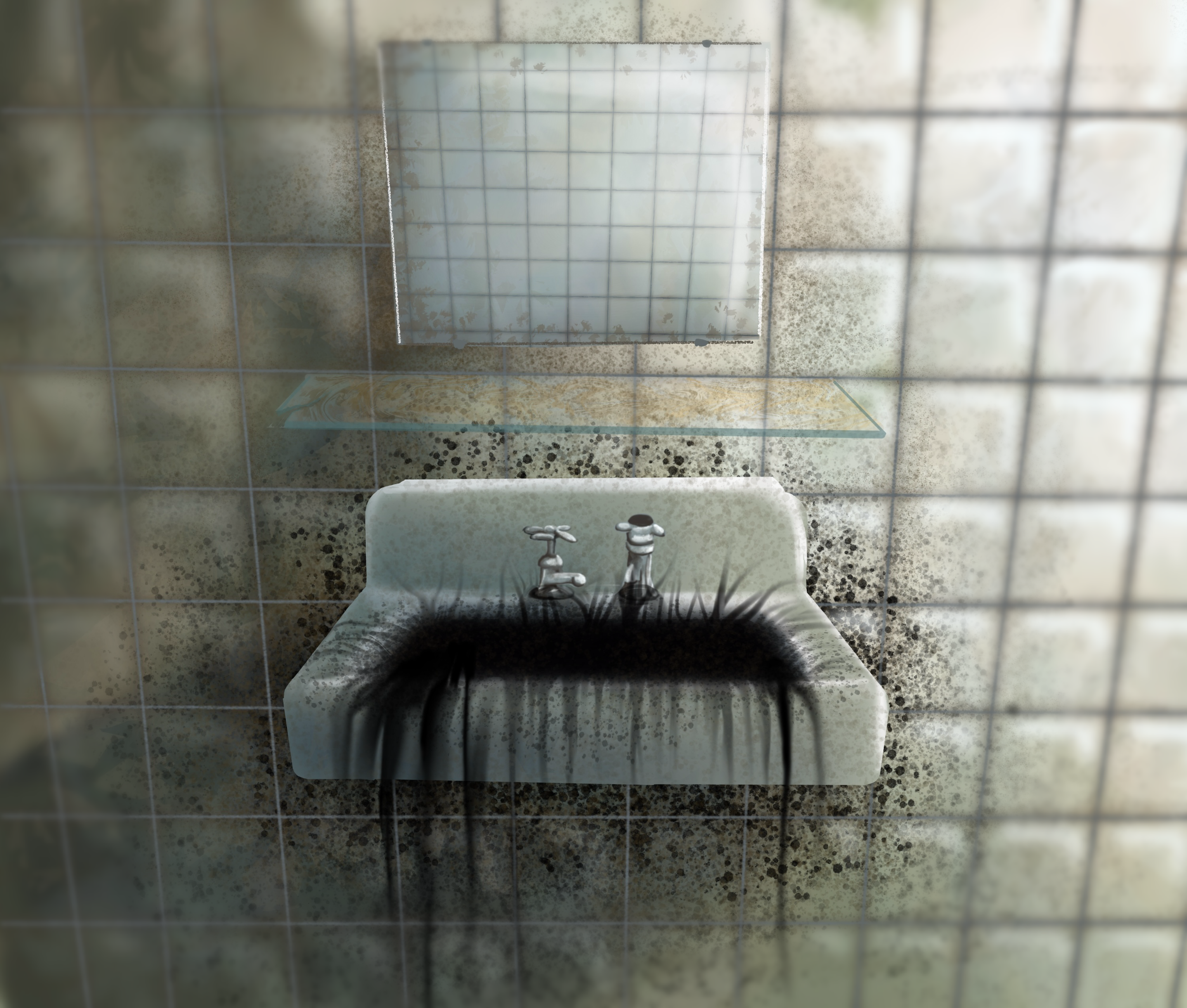
After a long day at work, you return home to find water pooling at the foot of your bathroom sink. Your heart sinks as you open the cabinet under the sink and find that a pipe has been slowly leaking. You quickly scramble to clean up the mess, but something feels odd as you start mopping the floor. There is a faint, musty smell in the air, something you can’t quite place. You think it’s just the dampness at first, but as time passes, the lingering smell grows stronger and more unsettling — until eventually it disappears. Then you start to see the dark green spots on the walls. You go from room to room, inspecting every nook and cranny, discovering more and more mold. What started as a slight inconvenience has become something much worse. Stachybotrys chartarum, known colloquially as black mold, is far more than just the dark-colored fungus that grows in the corners of your home [1]. Black mold exposure has been linked to various health problems, including fatigue, headache, persistent coughing, congestion, chest tightness, and loss of smell [2, 3, 4]. While it may be easy to dismiss a few black spots on your shower curtain, the insidious nature of black mold has the power to wreak havoc on your everyday life.
Mold Mine: What is Black Mold?
Black mold is one of the most common infectious fungi that grows indoors, distinguished from other household molds by its unique physical and chemical characteristics [4]. Though the fungus is named for its distinctive tar-like appearance, it can also appear green or dark brown, depending on its environmental conditions [3, 4]. The most crucial factors for black mold growth are high humidity and moisture, making areas affected by water infiltration, damage, or leaks ideal environments for its development [3]. Although black mold thrives in warmer temperatures, it can survive across a wide temperature range, from 36.5ºF to over 98ºF, allowing it to grow in diverse environments [3]. In order to survive, black mold feeds on cellulose, a sugar that provides structural support within plant cell walls [3, 5]. Drywall and wallpaper are examples of cellulose-rich materials found in homes that allow black mold to grow unseen [1, 6]. Think of the warm, moist environment ideal for mold growth like a middle school cafeteria: perfect for people to gather and for rumors to spread quickly. Similar to how pieces of information are overheard and passed between groups of students, black mold can diffuse through wallpaper and spread throughout an entire house, often going unnoticed until it is too late [6].
Spore War: Why You Can’t Smell
The small inconvenience that started in your bathroom took over your whole house through the specialized airborne reproductive cells of fungi called spores [7, 8]. Black mold spores can enter and harm the body via inhalation, ingestion, or direct contact [3, 9]. While one can avoid touching or ingesting moldy objects, it is much harder to protect oneself from inhaling airborne spores [3, 10]. With inhalation, black mold spores enter the body through the nose and release mycotoxins, which are small molecules produced by fungi that can pose a hazard to the health of other organisms [11, 12, 13]. The mycotoxins diffuse through the nasal epithelium, a protective tissue that lines the nasal cavity [9, 14, 15]. A large segment of the nasal epithelium, known as the olfactory epithelium, is located in the upper half of the nose and is responsible for detecting smells [16]. The olfactory epithelium contains olfactory sensory neurons that send signals to the olfactory bulb, the first part of the brain responsible for processing smell [17, 18]. The mycotoxins diffuse through the olfactory epithelium and use the olfactory sensory neurons to reach the olfactory bulb and subsequently the rest of the brain, wreaking all sorts of havoc [18, 19, 20].

Fuzzy Brain: Neurological Effects of Black Mold
Mycotoxins cause damage in essential bodily processes by disrupting the immune response and increasing inflammation [21, 22]. The most potent types of black mold mycotoxins are satratoxins, which increase the amount of free radicals in the brain [22, 23]. Free radicals are highly reactive molecules that can be neutralized by compounds called antioxidants. [24, 25] When free radicals vastly outnumber antioxidants, the overwhelming imbalance induces a destructive state that disrupts cellular function and communication referred to as oxidative stress [1, 3, 26]. The excess free radicals caused by satratoxins react with lipids, proteins, and DNA, and in the case of black mold mycotoxins, microglia [22, 24, 27, 28]. Microglia are one of the brain’s primary immune cells that play a key role in triggering neuroinflammation [29]. Inflammation is part of the body’s immune response against harmful stimuli, such as mycotoxins; when it occurs in the brain, it is known as neuroinflammation [28]. In oxidative stress, free radicals activate microglia, which in turn activate proteins called mitogen-activated protein kinases (MAPKs), increasing neuroinflammation and subsequently impacting neuronal function [27, 30, 31].
MAPKs cause two extremely damaging processes: inflammation and cell death [21, 32]. Inflammation is helpful in the short term as it helps the body respond to injury or infection; however, chronic inflammation can lead to tissue damage [28]. Inhaling the mycotoxin-containing spores from black mold over a long time leads to chronic inflammation and damages the surrounding nasal tissues [21]. Additionally, tissues are further damaged through cell death induced by the MAPKs [32]. The combination of cell death and neuroinflammation causes permanent damage to the body, especially in the olfactory system [21, 33]. A loss of smell stems from the death of olfactory sensory nerves as well as the atrophy of the olfactory epithelium and olfactory bulb [4, 33]. Prolonged neuroinflammation associated with black mold infection may increase susceptibility to neurodegenerative and sinus disorders [21, 33]. One proposed treatment avenue for black mold infection is supplementing an antioxidant known as glutathione [21]. Since glutathione deficiency is associated with the oxidative stress caused by black mold toxins, supplementing glutathione could neutralize the effects of satratoxins [21, 34]. Nasally administered glutathione has been found to reduce mycotoxin-induced neurocognitive symptoms [21]. Timely recognition and intervention are vital to mitigating the health risks associated with mold exposure and preventing further complications [35, 36].
Mold Cold: Why You’re Sick at Work
Black mold has been attributed to a neurological disease known as sick building syndrome (SBS) [37]. A term first coined in the 1970s, SBS refers to a series of nonspecific respiratory and neurological symptoms experienced by a group of residents or coworkers who spend most of their time in a particular building [38]. Headache, fatigue, difficulty concentrating, dizziness, nausea, and irritation of the eyes, nose, and throat are common symptoms of SBS [37]. Individuals affected by SBS often feel a sense of relief from their symptoms soon after leaving the building [37]. Many possible causes of SBS exist, which may vary from building to building [39]. One potential risk factor is poor ventilation, as pollutants can accumulate indoors and be inhaled by unknowing inhabitants when buildings are inadequately ventilated [38,39]. A number of pollutants have been associated with SBS, including chemicals like formaldehyde and biological material like animal droppings [37]. Notably, the growth of black mold is also a prominent cause of SBS [37]. There is an association between the presence of indoor mold and SBS symptoms; visible mold has also been linked to increased SBS symptoms [40]. Targeting the source of the pollutants is the usual treatment for SBS, such as removing air ventilation filters, replacing water-damaged ceiling tiles and carpeting, and installing new filters to clean the air [37].

Don’t Let Mold Take Hold: Avenues for Future Treatments
The discussion of black mold as a cause of neurological issues is relatively new, and further research is needed to understand how black mold affects the body [41]. The complex nature of mold exposure, individual variability in responses, and challenges with isolating mold-specific effects are some limiting factors regarding treatment [42]. Antioxidants, like glutathione, have been proposed as treatments because of their ability to counter oxidative stress and reduce neuroinflammation [21]. But these treatments are still in their infancy [21]. Some individuals who are experiencing symptoms from mold exposure have instead turned to unconventional treatments, including changing their diets, taking antifungal medications, and undergoing detoxification processes [36]. However, unconventional therapies tend to lack evidence of effectiveness [36]. Although many treatments have been proposed to combat satratoxins and their damaging effects, the most reliable remedy is prevention of exposure [36]. A common thread among proposed solutions is the need to either eliminate the mold or remove oneself from the contaminated environment, whether by arranging a mold inspection of the living space or taking the opportunity to move [41]. Relocating, however, can present challenges in many situations, reinforcing the importance of early recognition to prevent the spread of black mold. Though research is scarce, every day we are learning more about black mold: how it spreads, how it damages the brain, and how it can be treated.
Reference List
Tribelhorn, K., Twarużek, M., Kosicki, R., Straubinger, R. K., Ebel, F., & Ulrich, S. (2023). A chemically defined medium that supports mycotoxin production by Stachybotrys chartarum enabled analysis of the impact of nitrogen and carbon sources on the biosynthesis of macrocyclic trichothecenes and stachybotrylactam. Applied and Environmental Microbiology, 89(7). doi:10.1128/aem.00163-23
Lemons, A. R., Croston, T. L., Goldsmith, W. T., Barnes, M. A., Jaderson, M. A., Park, J. H., McKinney, W., Beezhold, D. H., & Green, B. J. (2019). Cultivation and aerosolization of Stachybotrys chartarum for modeling pulmonary inhalation exposure. International Forum for Respiratory Research, 31(13-14), 446-456.
Dyląg, M., Spychała, K., Zielinski, J., Łagowski, D., & Gnat, S. (2022). Update on Stachybotrys chartarum—Black mold perceived as toxigenic and potentially pathogenic to humans. Biology, 11(3), 352. doi:10.3390/biology11030352
Ibrahim, S. R. M., Choudhry, H., Asseri, A. H., Elfaky, M. A., Mohamed, S. G. A. & Mohamed, G. A. (2022). Stachybotrys chartarum—A hidden treasure: Secondary metabolites, bioactivities, and biotechnical relevance. Journal of Fungi, 8(5), 504. doi:10.3390/jof8050504
Höfte, H., & Voxeur, A. (2017). Plant cell walls. Current Biology, 27(17), R865-R870. doi:10.1016/j.cub.2017.05.025
Vance, P. H., Schaeffer, F., Terry, P., Trevino, E., & Weissfeld, A. S. (2016). Mold causes and effects “in a material world”. Clinical Microbiology Newsletter, 38(14), 111-116. doi:10.1016/j.clinmicnews.2016.06.004
Bava, R., Castagna, F., Piras, C., Musolino, V., Lupia, C., Palma, E., Britti, D., & Musella, V. (2022). Entomopathogenic fungi for pests and predators control in beekeeping. Veterinary Sciences, 9(2), 95. doi:10.3390/vetsci9020095
Oneto, D. L., Golan, J., Mazzino, A., Pringle, A., & Seminara, A. (2020). Timing of fungal spore release dictates survival during atmospheric transport. Proceedings of the National Academy of Sciences, 117(50), 5134-5143. doi:10.1073/pnas.1913752117
Kraft, S., Buchenauer, L., & Polte, T. (2021). Mold, mycotoxins, and a dysregulated immune system: A combination of concern? International Journal of Molecular Sciences, 22(22). doi:10.3390/ijms222212269
Croston, T. L., Lemons, A. R., Barnes, M. A., Goldsmith, W. T., Orandle, M. S., Nayak, A. P., Germolec, D. R., Green, B. J., & Beezhold, D. H. (2020). Inhalation of Stachybotrys chartarum fragments induces pulmonary arterial remodeling. American Journal of Respiratory Cell and Molecular Biology, 62(5), 563-576.
Omotayo, O. P., Omotayo, A. O., Mwanza, M., & Babalola, O. O. (2018). Prevalence of mycotoxins and their consequences on human health. Toxicological Research, 35(1), 1-7. doi:10.5487/TR.2019.35.1.001
Zhao, Z., Zhang, Z., Zhang, H., & Liang, Z. (2022). Small peptides in the detection of mycotoxins and their potential applications in mycotoxin removal. Toxins, 14(11), 795. doi:10.3390/toxins14110795
McCarty, L. S., Borgert, C. J., & Burgoon, L. D. (2020). Evaluation of the inherent toxicity concept in environmental toxicology and risk assessment. Environmental Toxicology and Chemistry, 39(12), 2351-2360. doi:10.1002/etc.4881
Marcelloni, A. M., Pigini, D., Chiominto, A., Gioffrè, A., & Paba, E. (2023). Exposure to airborne mycotoxins: The riskiest working environments and tasks. Annals of World Exposures and Health, 68(1), 19-35. doi:10.1093/annweh/wxad070
Scherzad, A., Hagen, R., & Hackenberg, S. (2019). Current understanding of nasal epithelial cell mis-differentiation. Journal of Inflammation Research, 12, 309-317. doi:10.2147/JIR.S180853
Choi, R., & Goldstein, B. J. (2018). Olfactory epithelium: Cells, clinical disorders, and insights from an adult stem cell niche. Laryngoscope Investigative Otolaryngology, 3(1), 35-42. doi:10.1002/lio2.135
Middleton, S. J., Perez-Sanchez, J., & Dawes, J. M. (2022). The structure of sensory afferent compartments in health and disease. Journal of Anatomy, 241(5), 1186-1210. doi:10.1111/joa.13544
Omura, K., Han, B., Nishijima, H., Aoki, S., Ebihara, T., Kondo, K., Otori, N., Kojima, H., Yamasoba, T., & Kikuta, S. (2021). Heterogeneous distribution of mature olfactory sensory neurons in human olfactory epithelium. International Forum of Allergy & Rhinology, 12(3), 266-277. doi:10.1002/alr.22885
Hiranuma, M., Okuda, Y., Fujii, Y., Richard, J. P., & Watanabe, T. (2024). Characterization of human iPSC-derived sensory neurons and their functional assessment using multi electrode array. Scientific Reports, 14. doi:10.1038/s41598-024-55602-8
Ehsanifar, M., Rajati, R., Gholami, A., & reiss, J. P. (2023). Mold and mycotoxin exposure and brain disorders. Journal of Integrative Neuroscience, 22(6), 137. doi:10.31083/j.jin2206137
Chaudhary, Z., Rehman, K., & Akash M. S. H. (2021). Mechanistic insight of myotoxin-induced neurological disorders and treatment strategies. In: Akash, M. S. H., & Rehman, K. (eds.) Environmental Contaminants and Neurological Disorders. Emerging Contaminants and Associated Treatment Technologies. Springer. doi:10.1007/978-3-030-66376-6_7
Mavrommatis, A., Giamouri, E., Tavrizelou, S., Zacharioudaki, M., Danezis, G., Simitzis, P. E., Zoidis, E., Tsiplakou, E., Pappas, A. C., Georgiou, C. A., & Feggeros, K. (2021). Impact of mycotoxins on animals' oxidative status. Antioxidants, 10(2). doi:10.3390/antiox10020214
Cleary, D. (2022). Effects of mycotoxins on macrophages and their possible clinical implications. In: Gupta. S., & Pathak, Y. V. (eds.) Macrophage Targeted Delivery Systems. Springer. doi:10.1007/978-3-030-84164-5_23
Chandimali, N., Bak, S. G., Park, E. H., Lim, H. J., Won, Y. S., Kim, E. K., Park, S. I., & Lee, S. J. (2025). Free radicals and their impact on health and antioxidant defenses: A review. Cell Death Discovery, 11. doi:10.1038/s41420-024-02278-8
Tan, B. L., Norhaizan, M. E., Liew, W. P. P., & Rahman, H. S. (2018). Antioxidant and oxidative stress: A mutual interplay in age-related diseases. Frontiers in Pharmacology, 9. doi:10.3389/fphar.2018.01162
Hassan, H. A., Ahmed, H. S., & Hassan, D. F. (2024). Free radicals and oxidative stress: Mechanisms and therapeutic targets. Human antibodies, 32(4), 151-167. doi:10.3233/HAB-240011
Kumar, R. R., Arora, K., Goswami, S., Sakhare, A., Singh, B., Chinnusamy, V., & Praveen, S. (2020). MAPK enzymes: A ROS activated signaling sensors involved in modulating heat stress response, tolerance and grain stability of wheat under stress. 3 Biotech, 10(9). doi:10.1007/s13205-020-02377-0
Chen, M. J., Ramesha, S., Weinstock, L. D., Gao, T., Ping, L., Xiao, H., Dammer, E. B., Duong, D. D., Levey, A. I., Lah, J. J., Seyfried, N. T., Wood, L. B., & Rangaraju, S. (2021). Inflammatory responses and inflammation-associated diseases in organs. Journal of Neuroscience Research, 99(6), 1704-1721. doi:10.1002/jnr.24829
Shao, F., Wang, X., Wu, H., Wu, Q., & Zhang, J. (2022). Microglia and neuroinflammation: Crucial pathological mechanisms in traumatic brain injury-induced neurodegeneration. Frontiers in Aging Neuroscience, 14. doi:10.3389/fnagi.2022.825086
Ferro, A., Aguste, Y. S., S., & Cheadle, L. (2021). Microglia, cytokines, and neural activity: Unexpected interactions in brain development and function. Frontiers in Immunology, 12. doi:10.3389/fimmu.2021.703527
Ishihara, Y., Itoh, K. (2022). Microglial inflammatory reactions regulated by oxidative stress. Journal of Clinical Biochemistry and Nutrition, 72(1), 23-27. doi:10.3164/jcbn.22-71
Vervaeke, A., & Lamkanfi, M. (2025). MAP kinase signaling at the crossroads of inflammasome activation. Immunological reviews, 329(1). doi:10.1111/imr.13436
LaFever, B. J., & Imamura, F. (2022). Effects of nasal inflammation on the olfactory bulb. Journal of Neuroinflammation, 9(19). doi:10.1186/s12974-022-02657-x
Kulcsár, S., Kövesi, B., Balogh, K., Zándoki, E., Ancsin, Z., Erdélyi, M. & Mézes, M. (2023). The co-occurrence of T-2 toxin, deoxynivalenol, and fumonisin B1 activated the glutathione redox system in the EU-limiting doses in laying hens. Toxins, 15(5). doi:10.3390/toxins15050305
Hurraß, J., Nowak, D., Heinzow, B., Joest, M., Stemler, J., & Wiesmüller, G. A. (2024). Indoor mold. Deutches Ärtzeblatt, 121, 265-271. doi:10.3238/arztebl.m2024.0018
Rea, W. J. (2018). A large case-series of successful treatment of patients exposed to mold and mycotoxin. Clinical Therapeutics, 40(6), 889-893. doi:10.1016/j.clinthera.2018.05.003
Nag, P. K. (2018): Sick building syndrome and other building-related illnesses. Office Buildings, 53-103. doi:10.1007/978-981-13-2577-9_3
Subri, M. S. M., Arifin, K., Sohaimin, M. F. A. M., & Abas, A. (2024). The parameter of the sick building syndrome: A systematic literature review. Heliyon, 10(12). doi:10.1016/j.heliyon.2024.e32431
Mansor, A. A., Abdullah, S., Ahmad, A. N., Ahmed, A. N., Zulkifli, M. F. R., Jusoh, S. M., & Ismail, M. (2024). Indoor air quality and sick building syndrome symptoms in administrative office at public university. Dialogues in Health, 4. doi:10.1016/j.dialog.2024.100178
Lu, C., Deng, Q., Li, Y., Sundell, J., & Norbäck, D. (2016). Outdoor air pollution, meteorological conditions and indoor factors in dwellings in relation to sick building syndrome (SBS) among adults in China. Science of the Total Environment, 560-561, 186–196. doi:10.1016/j.scitotenv.2016.04.033
U.S. Department of Health and Human Services National Toxicology Program (2024). NTP technical report on the toxicity study of Stachybotrys chartarum (CASRN 67892-26-6) administered by inhalation to B6C3F1/N mice. Retrieved from https://ntp.niehs.nih.gov/sites/default/files/2024-09/tox107_508.pdf
Harding, C. Ff., Pytte, C. L., Page, K. G., Ryberg, K. J., Normand, E., Remigio, G. J., DeStefano, R. A., Morris, D. B., Voronina, J., Lopez, A., Stalbow, L. A., Williams, E. P., & Abreu, H. (2021). Mold inhalation causes innate immune activation, neural, cognitive, and emotional dysfunction. Brain, Behavior, and Immunity, 87, 218-228. doi:10.1016/j.bbi.2019.11.006